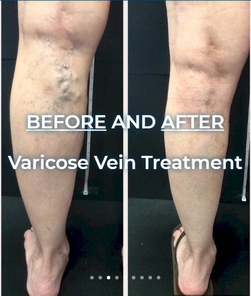
Before any treatment plan is made, varicose veins need to be properly diagnosed. The visible bulging veins on the surface are often only part of the picture — the real source of the problem usually sits deeper in the leg, where faulty valves allow blood to pool. The way we find that source is with a painless test called a duplex ultrasound, often referred to as vein mapping. Understanding how the diagnosis works can take a lot of the uncertainty out of your first visit.
Why Diagnosis Starts With an Ultrasound, Not Just a Look
Looking at the legs only tells part of the story. Two people can have very similar-looking veins on the surface but completely different underlying causes. A duplex ultrasound combines a picture of the vein with a reading of how blood moves through it, so we can see which valves have weakened and whether blood is flowing backward (a condition called venous reflux). This is why an accurate ultrasound vein diagnosis matters: it guides every decision that follows, from whether treatment is needed at all to which approach fits your anatomy.
What Ultrasound Vein Mapping Actually Shows
Vein mapping is a careful, methodical scan of the veins in the leg. It is usually done while you are standing or sitting, because gravity makes valve problems easier to detect. During the scan we look for several things:
- Which veins are affected and how far the problem extends
- Whether blood is refluxing (flowing the wrong direction) and for how long
- The size and depth of the veins involved
- Any blood clots or blockages that need to be ruled out
- A clear “map” that shows the exact points to target during treatment
This map is what turns a general complaint — aching, heaviness, swelling, or visible vein symptoms — into a specific, treatable picture.
What to Expect During the Test
The scan itself is comfortable and non-invasive. There are no needles and no recovery time. A technician or physician applies a small amount of gel and moves a handheld probe along the leg while watching the images on screen. Most mappings take roughly 30 to 45 minutes per leg. You can drive yourself home and return to normal activity right away. The findings are then reviewed so your options can be explained in plain language.
How the Diagnosis Shapes Your Treatment Plan
Once mapping is complete, the results point toward the right approach for your situation. Depending on what the ultrasound reveals, options may include varicose vein treatment such as endovenous laser (EVLA) to close a faulty vein, Varithena foam treatment, or VenaSeal closure. In some cases, conservative steps like compression are recommended first. Because the diagnosis is specific to your veins, the plan is too — there is no one-size-fits-all answer.
A Note on Insurance and Cost
Most PPO plans and Medicare cover treatment for symptomatic veins, which means the diagnostic ultrasound and any medically necessary treatment that follows are often covered when symptoms are present. Purely cosmetic vein work is self-pay. We can review your coverage with you before anything is scheduled.
Care Backed by Experience
At our practice, diagnosis is handled with the same care as treatment. Dr. Abboud brings more than 20 years of experience to every evaluation. He is a graduate of the University of Damascus and completed his residency at Cook County Hospital and John H. Stroger Jr. Hospital in Chicago. You can learn more about Dr. Abboud and how we approach vein care across our three locations: Chicago (5907 W. 63rd St.), Oak Brook (2425 W. 22nd St. #205), and Orland Park (10 Orland Square Dr.).
If you have been wondering what is really going on beneath the surface, the first step is a clear diagnosis. Book a Free Vein Screening or call us at 630-974-1400, and we will walk you through what your ultrasound shows and what your options are.